
Health
How to Prevent Asthenia

HOW TO PREVENT ASTHENIA
How to prevent asthenia. Body weakness, or asthenia, can lead to physical, depression and occasionally mental exhaustion. Lack of energy, twitchy muscles, or cramping make it hard or impossible to move your body. A person may have asthenia in one or more body parts, like the arms or legs. Others might suffer from full-body weakness, which is frequently brought on by a viral or bacterial infection like influenza or hepatitis.. Sometimes weakness is chronic or ongoing, but it can also be transient. Find out more about asthenia’s potential symptoms, causes, and when to consult your doctor.
HOW TO PREVENT ASTHENIA
Usually, underlying medical disorders are the cause. Acute causes include infections, which can cause muscle stiffness, and cardiac decompensation, which can result from a heart attack or stroke. Natural aging, malnutrition, nutritional imbalances (such as a lack of vitamin B-12), anemia, hypothyroidism, diabetes, TB, sleep apnea, and mental health issues including depression are examples of chronic causes.
Symptoms
A single vulnerability
You can discover that you are unable to move a certain body part effectively if you experience weakness in that location. Additionally, you might encounter: • Slow or delayed motion • Uncontrollable tremors or shaking • twitching of muscles • cramping in the muscles weakness throughout the body You feel exhausted, much like you do when you have the flu, due to whole body weakness. Although this is referred to as weariness, full-body weakness can also occur without exhaustion. Some individuals who suffer from weakness across their entire body also have:
Prevention

1. flu-like symptoms and fever
2. discomfort in the afflicted area Symptoms of an emergency If you encounter any of the following signs, you ought to speak with your physician.
3. lightheadedness, dizziness, and confusion Speaking difficulties, eyesight problems, chest pain, and breathing difficulties
Causes
Treating the underlying cause may be the most effective strategy to prevent asthenia because it is linked to a number of different illnesses. Other potential preventive actions include consuming a healthy diet and exercising.
• Addressing underlying sleep challenges
• using prescription drugs, if necessary.
• taking iron, folate, or vitamin B12 supplements, if you are lacking in any of these nutrients.
• cutting back on alcohol and caffeine
Cancer

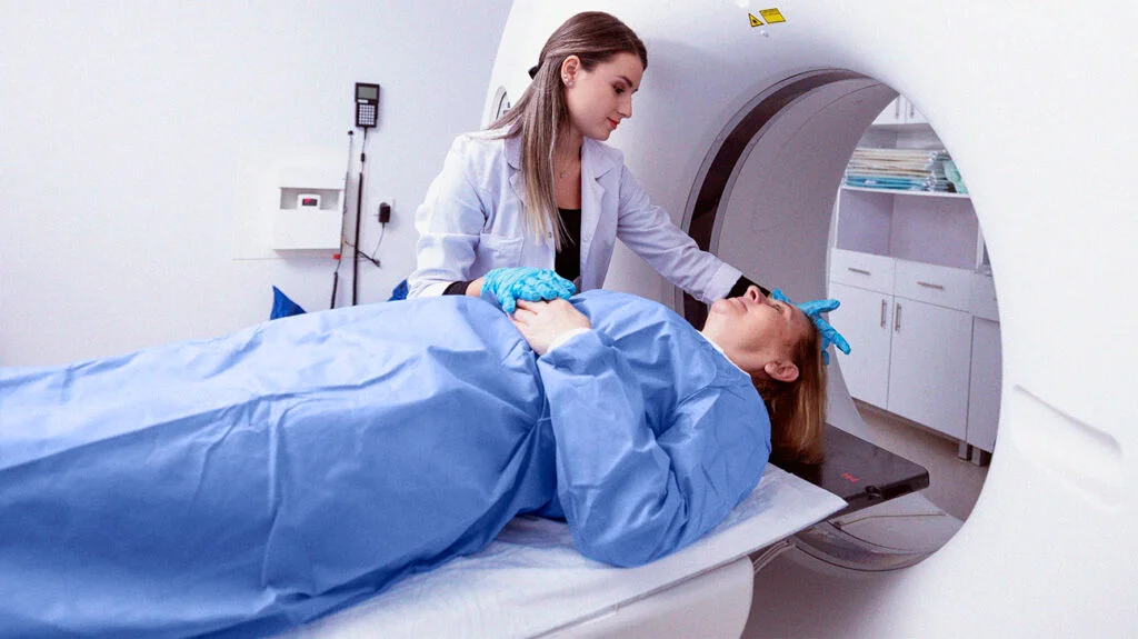
Your doctor will go over your treatment options if cancer is the reason for your frailty. The optimum course of treatment is determined in part by the body structure, stage, and location. Among the cancer treatment choices are chemotherapy and radiation therapy. • Surgery Asthenia can also result after chemotherapy and other cancer treatments.
Dehydration

However, increasing your hydration intake can help if you’re dehydrated. You could need medical treatment, though, if your dehydration symptoms are severe. You will be given fluids via an intravenous line in the hospital. In order to raise your blood pressure, you could also require medication. At this stage, the weakness might start to lessen.
Anemia
Meanwhile, if anemia is the cause of your weakness, you might require iron supplements if you seem to be iron deficient. If your anemia is severe, you might require a blood transfusion. The hospital will give you a blood transfusion if you require one. An IV line is used to receive donor blood as part of this treatment.
A heart attack
Also, your doctor will talk to you about your treatment options if your weakness was brought on by a heart attack. Not every instance of weakness needs to be treated. Treatment might not be required if your weakness is the result of the flu or a cold.
Summary
Extreme weakness and exhaustion are generally referred to as asthenia, however there are many different and sometimes complicated underlying causes. This disorder can cause significant physical or mental weakness that can be linked to a more serious emergency, a long-term medical condition, or a short-term illness.


Remedies for breast cancer. Alongside these medication interventions, lifestyle changes including eating a balanced diet, exercising frequently, and abstaining from alcohol and tobacco are also important. Additionally, stress management can help with both general wellbeing and breast cancer treatment. People who have been diagnosed with breast cancer should collaborate closely with their medical team. to create a thorough treatment strategy that takes into account their particular requirements and objectives.
REMEDIES FOR BREAST CANCER
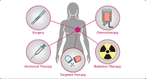
Breast cancer is a disease in which the breast’s cells proliferate uncontrollably, creating tumors that may spread and become invasive. Although it can happen to younger women and men, it mainly affects women over 50. A new lump, breast thickening, skin dimpling, or nipple discharge are important indicators. Treatment options include radiation, chemotherapy, hormone therapy, and surgery, all of which have a high success rate.
1. Surgery:

Surgery is often the first line of treatment for breasts cancer. The type of surgery depends on the size and location of the tumor, as well as other factors. Common surgical options include lumpectomy (removal of the tumor and a small margin of surrounding tissue) and mastectomy (removal of the entire breast). In some cases, lymph nodes may also be removed to determine if cancer has spread.
2. Chemotherapy:
Chemotherapy involves the use of powerful drugs to destroy cancer cells or shrink tumors. It may be used before surgery (neoadjuvant chemotherapy) to reduce the size of the tumor, after surgery (adjuvant chemotherapy) to kill any remaining cancer cells, or as the primary treatment for advanced or metastatic breasts cancer.
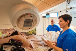
3. Radiation Therapy:
Remedies for breast cancer
Radiation therapy uses high-energy beams to kill cancer cells or prevent them from growing. It is often used after surgery to destroy any remaining cancer cells in the breast, chest wall, or lymph nodes. Radiation therapy may also be used to relieve symptoms in cases of advanced breast cancer.
4. Hormone Therapy:
Hormone therapy, also known as endocrine therapy, targets hormone receptors on cancer cells that rely on estrogen and/or progesterone to grow. Hormone therapy may include medications such as tamoxifen, aromatase inhibitors, or ovarian suppression therapy to block the effects of estrogen or reduce its production.
5. Targeted Therapy:
Remedies for breast cancer
Targeted therapy drugs specifically target cancer cells while sparing healthy cells. Examples of targeted therapy drugs used in breast cancer treatment include trastuzumab (Herceptin) and pertuzumab (Perjeta), which target HER2-positive breasts cancer, and CDK4/6 inhibitors such as palbociclib, ribociclib, and abemaciclib, which target certain proteins involved in cell growth.
6. Immunotherapy:
Also, immunotherapy works by stimulating the body’s immune system to recognize and attack cancer cells. While not yet widely used in breast cancer treatment, immunotherapy drugs such as pembrolizumab and atezolizumab may be used in certain cases, particularly in metastatic breasts cancer that is triple-negative.
7. Clinical Trials:

Remedies for breast cancer
Clinical trials offer access to new and experimental treatments for breast cancer. Participation in clinical trials helps advance medical knowledge and may provide patients with access to promising therapies not yet available to the general public.
Summary
A variety of medical interventions that are particular to the kind and stage of the cancer are usually used to treat breast cancer. Medical professionals stress that clinical therapies are the main means of targeting and eradicating cancer cells, despite the fact that “remedies” frequently refer to home remedies.
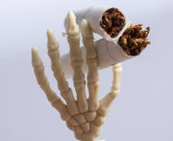
General health and smoking. Inhaling smoke from burning materials, mostly tobacco, through pipes, cigars, or cigarettes is known as smoking. Nicotine, which provides momentary pleasure and relaxation but causes serious health problems like cancer, heart disease, stroke, and lung illness, makes it a highly addictive habit.
GENERAL HEALTH AND SMOKING
Smoking damages almost every organ in the body and lowers general health, making it the biggest cause of avoidable disease and death worldwide. Every year, it results in over 7 million deaths and an average 10-year reduction in life expectancy. It affects almost all of the body’s organs and has disastrous repercussions on health. The following are a few of the most serious health effects of smoking:
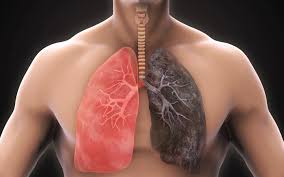
1. Respiratory System:
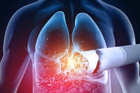
Smoking damages the lungs and airways, leading to chronic conditions like chronic obstructive pulmonary disease (COPD), emphysema, and chronic bronchitis. It also increases the risk of respiratory infections like pneumonia and xacerbates asthma symptoms.
2. Cardiovascular System: Smoking significantly increases the risk of heart disease, stroke, and peripheral artery disease. It damages the blood vessels, leading to atherosclerosis (hardening and narrowing of the arteries), which can cause heart attacks and strokes.
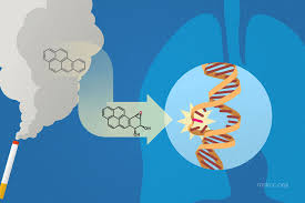
3. Cancer:
General health and smoking
Smoking is the leading cause of preventable cancer worldwide. It increases the risk of developing various types of cancer, including lung cancer, throat cancer, mouth cancer, esophageal cance, bladder cancer, pancreatic cancer, and cervical cancer.
4. Reproductive System:
Smoking harms reproductive health in both men and women. It reduces fertility, increases the risk of erectile dysfunction in men, and can lead to complications during pregnancy, such as miscarriage, preterm birth, low birth weight, and birth defects.
5. Immune System:
General health and smoking
Smoke weakens the immune system, making smokers more susceptible to infections and slower to heal from injuries and illnesses. It also increases the risk of autoimmune diseases and exacerbates existing autoimmune conditions.
6. Vision:
Smokers are associated with an increased risk of vision problems and eye diseases, including age-related macular degeneration (AMD), cataracts, and diabetic retinopathy.
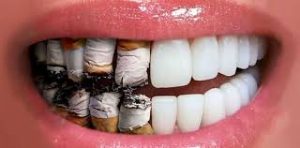
7. Oral Health:
General health and smoking
Smoking damages oral tissues, leading to gum disease, tooth loss, and oral cancer. It also causes bad breath, tooth discoloration, and a decreased sense of taste and smell.
8. Skin: It accelerates skin aging and increases the risk of skin conditions such as psoriasis and skin cancer. It also reduces blood flow to the skin, resulting in a dull complexion and delayed wound healing.
9. Bone Health:
It weakens bones and increases the risk of osteoporosis, a condition characterized by brittle and fragile bones, which is more common in older adults.
Summary
Overall, the health effects of smoking are extensive and profound, affecting not only smokers but also those exposed to secondhand smoke. Quitting smoking is the single most important step smokers can take to improve their health and reduce their risk of developing smoking-related diseases.
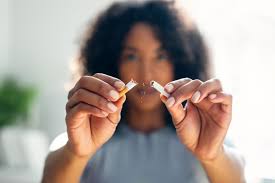
Tips for quitting smoking habits. Over 80% of the 1.3 billion smokers worldwide reside in low- and middle-income nations, and smoking habits are frequently motivated by nicotine dependence. Tobacco use becomes a deeply embedded ritual connected to stress reduction, social contact, and everyday routines. These behaviors, which include smoking 500–600 times a day, increase the risk of serious illnesses like cancer, lung infections, and cardiovascular disease.
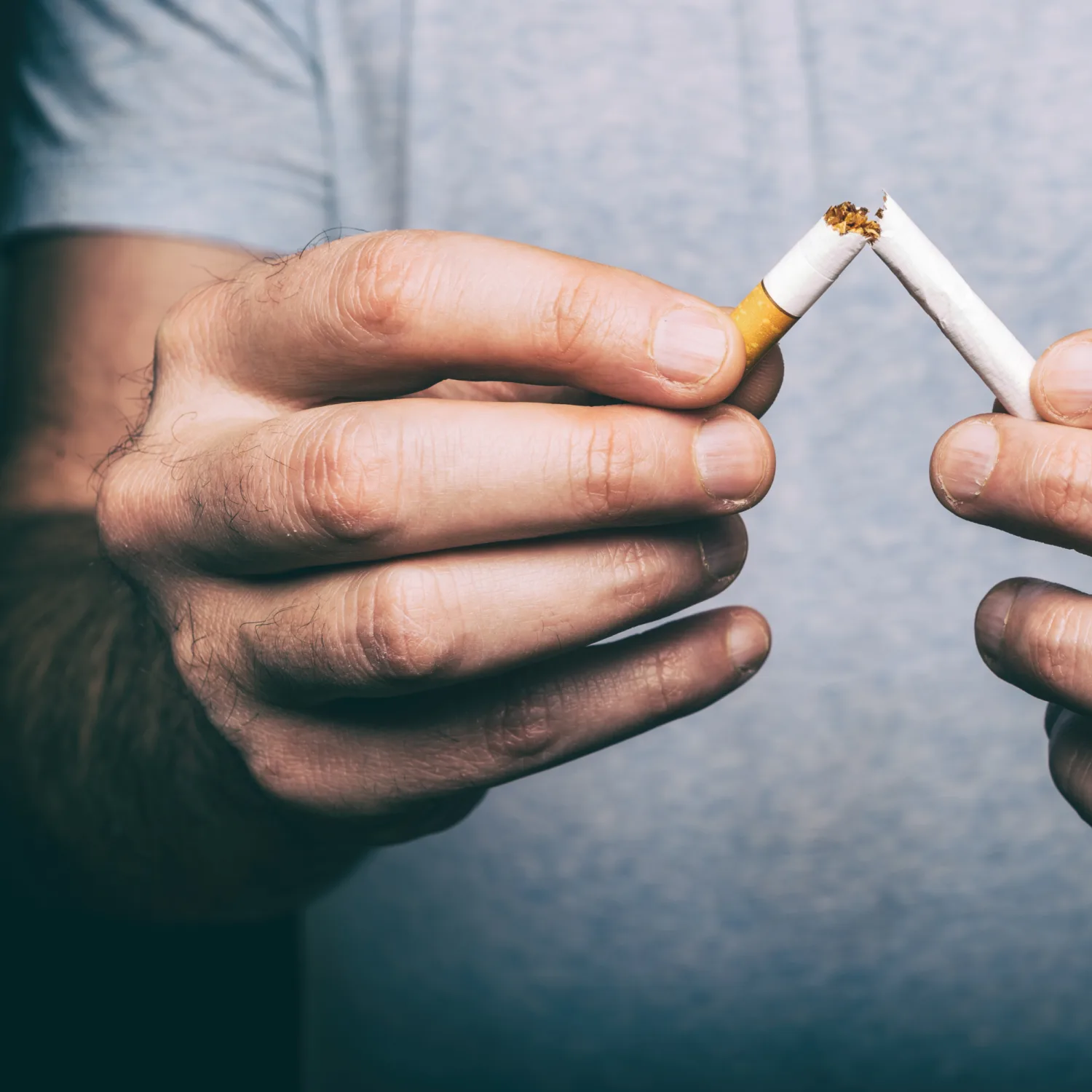
TIPS FOR QUITTING SMOKING HABITS
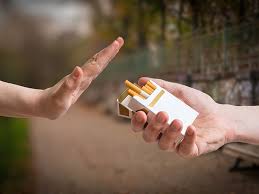
It’s never too late to start quitting smoking, which is one of the healthiest things you can do for your health. You can overcome the hold of smoking with perseverance and encouragement. Although quitting smoking can be difficult, it is undoubtedly possible with the correct strategy. Here’s a strategy to assist:
1. Set a Quit Date:
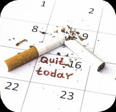
Choose a date within the next few weeks to quit smoking. This gives you time to prepare mentally and emotionally.
2. Understand Your Triggers: Identify situations, emotions, or activities that trigger your urge to smoke. This awareness will help you develop strategies to cope with these triggers.
3. Find Alternatives:
Replace smoking with healthier habits like chewing gum, taking a walk, or deep breathing exercises to distract yourself when cravings hit.
4. Seek Support: Tell your friends, family, and coworkers about your decision to quit smoking. Their support can make a big difference in your journey.
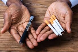
5. Consider Nicotine Replacement Therapy (NRT): NRT products like patches, gum, or lozenges can help ease withdrawal symptoms by providing a controlled dose of nicotine.
6. Stay Busy:

Tips for quitting smoking habits
Keep yourself occupied with activities that keep your hands and mind busy. This can reduce the urge to smoke.
7. Avoid Triggers: Stay away from situations or places where you’re tempted to smoke, especially during the early stages of quitting.
8. Stay Positive:
Tips for quitting smoking habits
Remind yourself why you want to quit smoking. Focus on the benefits of a smoke-free life, such as improved health, more energy, and saving money.
9. Be Patient and Persistent: Quitting smoking is a process, and setbacks may happen. Don’t get discouraged. Learn from any slip-ups and keep moving forward.
10. Celebrate Milestones:
Tips for quitting smoking habits
Reward yourself for reaching milestones along the way, whether it’s a week, a month, or a year without smoking. Treat yourself to something you enjoy as a reminder of your progress.
Summary
When behavioral modifications, nicotine replacement therapy (NRT), and social support are combined, quitting smoking is quite successful. Setting a specific stop date, recognizing and avoiding triggers, controlling cravings with nutritious snacks or diversions, and making use of support services like hotlines or counseling are all important stages.

Skin cancer remedies

Skin cancer preventive measures

Remedies for breast cancer

A Step-by-Step Guide to Deleting Reels on Instagram

A Comprehensive Guide to Setting Up a YouTube Premiere

How to Create a WhatsApp Group
-

 Music4 weeks ago
Lyrics for Iké Nilé
-

 Photography4 weeks ago
Photography and Outdoors
-

 Social media4 weeks ago
Tips for likes and Followers on Facebook
-

 Social media4 weeks ago
Guidelines for Creating a Facebook Page
-

 Business3 weeks ago
Business plan advantages
-

 Social media2 weeks ago
Tips for editing whatsapp messages