Health
Causes of Nosebleeds
CAUSES OF NOSEBLEEDS
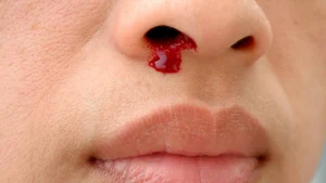
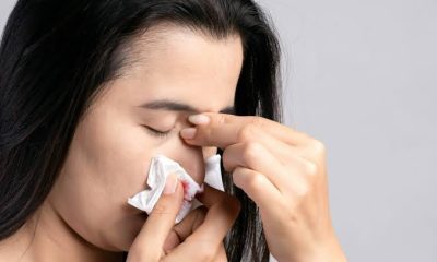
Causes of nosebleeds. A nosebleed occurs when a blood vessel in the nasal lining ruptures. Various factors can lead to nosebleeds, including infections, injuries, allergic reactions, nasal picking, or the insertion of foreign objects into the nostril. The medical term for a nosebleed is epistaxis. Nosebleeds are frequently seen in children and are generally not a cause for concern.
CAUSES OF NOSEBLEEDS
However, it is advisable to seek medical assistance if the nosebleeds are severe, recurrent, or last for an extended period. The delicate blood vessels located in the septum, the rigid tissue that separates the nostrils and divides the nose into two sections, are prone to rupture, which can lead to a nosebleed. In children, these nosebleeds typically occur on one side only (unilateral). Most children eventually outgrow this issue. However, if the bleeding is excessive, lasts for an extended period, or does not cease with initial first aid efforts, it is advisable to seek medical attention at a doctor’s office or an emergency department.
Symptoms
The indicators of a nosebleed consist of:
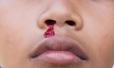
1. bleeding from one or both nostrils
2. feeling of liquid running down the throat
3. a frequent desire to swallow.
Causes
A nosebleed may result from various factors, such as:
delicate blood vessels that are prone to bleeding, particularly in warm, dry conditions or following physical activity
infections affecting the nasal lining, sinuses, or adenoids
allergic reactions leading to hay fever or persistent coughing
trauma from bumps or falls
the insertion of foreign objects into the nostril
habitual nose picking and, in some cases, issues related to bleeding or clotting disorders.
Treatment
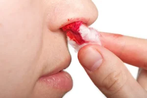
To effectively manage a nosebleed, follow these steps:
Provide reassurance to the individual, particularly children, as crying can exacerbate blood flow.
Have the person sit upright and tilt their head slightly forward.
Apply pressure with your fingers and thumb on the soft area of the nostrils just below the bridge of the nose for a minimum of 10 minutes.
Encourage the individual to breathe through their mouth while their nostrils are being pinched.
Loosen any tight clothing around the neck.
Place a cold cloth or ice pack on the person’s forehead and another around the neck, focusing on the sides.
After 10 minutes, release the pressure on the nostrils and check if the bleeding has ceased.
If the bleeding continues, seek medical assistance.
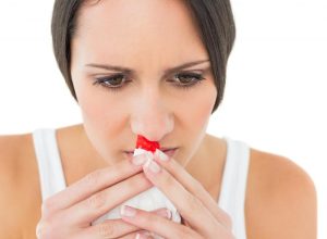
Instruct the individual to refrain from sniffing or blowing their nose for a minimum of 15 minutes, and to avoid picking their nose for the remainder of the day. The presence of clotted blood in the nasal passages can be uncomfortable, and children, in particular, may struggle to resist the urge to sniff or blow their nose for several hours. Allowing a period of 15 minutes will provide sufficient time for the clot to stabilize.
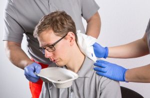
If the bleeding persists despite basic first aid measures, it is essential to seek medical attention from a doctor or visit a hospital emergency department. Identifying and addressing the underlying cause of the continued bleeding is crucial.
Summary
Anterior nosebleeds, which occur from blood vessels in the front section of the nose, can typically be managed at home by pinching the nostrils together for a continuous duration of 10 minutes while sitting upright. It is crucial to avoid pinching the bony upper region of the nose. The pinch should be firm and maintained without interruption throughout the entire 10 minutes. Alternative home remedies, such as applying ice packs to the nose, inserting tissue paper into the nostrils, or adjusting head positions, have proven to be ineffective.


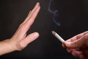
How to quit smoking. Smoking is the inhalation of smoke from burning tobacco (usually from pipes, cigars, or cigarettes), which spreads nicotine and more than 7,000 compounds throughout the body, including at least 69 recognized carcinogens. It damages almost every organ, especially the heart and lungs, and is the greatest preventable cause of death in the United States, accounting for around 480,000 deaths each year.
HOW TO QUIT SMOKING
Most tobacco users either want to smoke or have strong cravings for tobacco goods. But you can fight these urges. When you have a strong want to use tobacco, keep in mind that the need will likely go away in five to ten minutes, regardless of whether you indulge in a cigarette or a chewing tobacco dip. Every time you overcome a desire to smoke, you get one step closer to giving up tobacco use permanently.
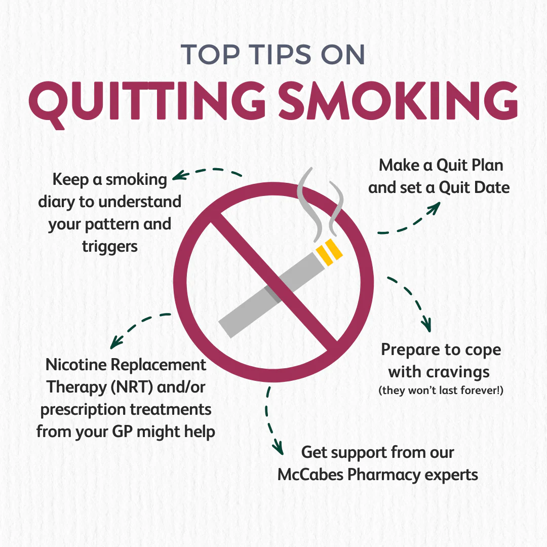
1. Consider utilizing nicotine replacement;
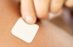
Inquire with your physician about nicotine replacement treatment. The available choices consist of Prescription nicotine in the form of an inhaler or nasal spray Over-the-counter nicotine patches, gum, and lozenges Prescription non-nicotine stop-smoking medications including bupropion
2. Avoid triggers;
Desires to smoke or chew tobacco are probably stronger in the settings where you used to smoke or chew tobacco, like bars or parties, or during stressful moments while you’re drinking coffee. Identify your triggers and make a plan to either avoid or deal with them.
3. Delay;

Tell yourself to hold off on giving in to your urge for tobacco if you feel like you need to wait ten minutes. Then, throughout that period, engage in some self-distraction.
4. Chew on it;
To help you avoid the need to smoke, give your mouth something to do. Enjoy chewing gum or hard candy. Alternatively, nibble on crunchy and delicious raw carrots, almonds, or sunflower seeds.
5. Don’t have ‘just one’;
How to quit smoking
If you feel a yearning for tobacco, you could be tempted to have just one cigarette. However, do not deceive yourself into believing that you should end there. Having one almost often results in having more. Additionally, you might start smoking again.
6. Get physical;
Engaging in physical activity can assist you in avoiding smoke cravings. A few quick movements, like rushing up and down the stairs, will help quell an urge for tobacco. Take a jog or a stroll outside.
7. Try relaxation techniques;

How to quit smoking
You might have used smoking as a stress-reduction strategy. It might be stressful to combat a tobacco urge on its own. Try these relaxation techniques to help you de-stress, like deep breathing, yoga, massage, muscular relaxation, visualization, and relaxing music.
Summary
Setting a specified “Quit Day,” using nicotine replacement therapies (patches, gum, lozenges) or prescription drugs (bupropion, varenicline), and altering daily routines to avoid triggers are the best ways to stop smoking. Cravings can be controlled with the support of friends, therapy, and methods like the “4 Ds” (Delay, Deep Breathe, Drink Water, Do Something Else).

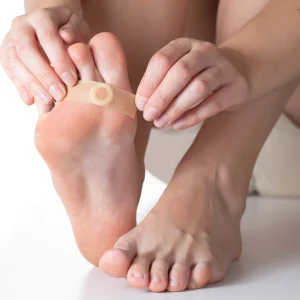
Calluses causes. Usually on the hands or feet, calluses are thickened, hardened, and frequently painless patches of skin brought on by constant pressure or friction, such as from wearing poorly fitted shoes, going barefoot, or using tools. Skin that is flaky, dry, or rough can be treated by soaking, lightly filing with a pumice stone, and wearing appropriate footwear. While consequences are uncommon but can include infection, particularly in people with diabetes or poor circulation, risk factors include wearing shoes that are excessively thin or going barefoot.
CALLUSES CAUSES
By decreasing friction and pressure on the skin through the use of orthotics, moisturizer, and well-fitting, cushioned shoes, calluses can be avoided. Wearing gloves when performing hard labor, utilizing pads (moleskin) to protect sensitive regions, and switching up footwear every day are important tactics. Additionally, regular, mild exfoliation with a pumice stone aids in preventing accumulation.
Causes of it;

Calluses often develop on the hands and feet, as these areas are most exposed to repetitive activities. Common causes include:
– Wearing tight or ill-fitting shoes.
– Walking barefoot or in high heels.
– Manual labor or activities like gardening, weightlifting, or playing musical instruments.
How to Prevent;

CALLUSES CAUSES
Prevention is key to avoiding discomfort. Here are some tips:
– Wear Proper Footwear: Ensure shoes fit well and provide adequate cushioning.
– Use Gloves: Protect your hands during activities that involve gripping tools or equipment.
– Moisturize Regularly: Keep your skin hydrated to prevent dryness and cracking.
Caring for Calluses;
CALLUSES CAUSES
If you already have calluses, these steps can help:
– Soak and Exfoliate: Soak the affected area in warm water to soften the skin, then gently scrub with a pumice stone.
– Apply Moisturizers: Use lotions containing urea or salicylic acid to soften the skin.
– Avoid Cutting: Do not attempt to cut or pick at calluses, as this can lead to infection.

CALLUSES CAUSES
If a callus becomes painful or shows signs of infection, consult a healthcare provider for treatment.
With proper care and attention, you can manage calluses and keep your skin healthy.
Summary
Thickened patches of skin known as calluses develop naturally as a result of constant pressure, friction, or discomfort. Although they are usually not dangerous, if addressed, they can be unpleasant and ugly.
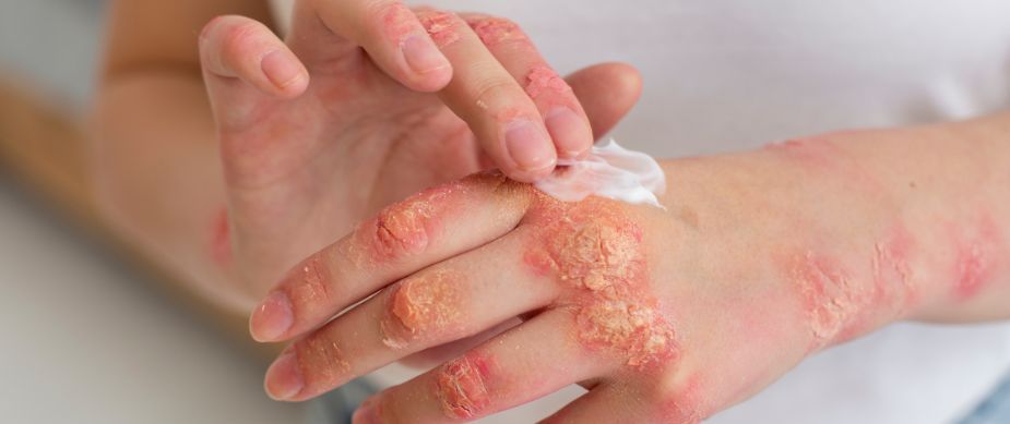
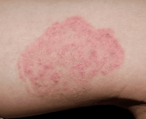
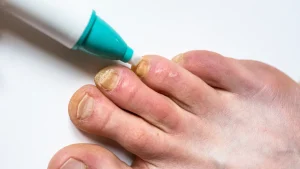
Signs of Fungal infections. Mycosis, another name for fungal infection, is a condition brought on by fungi. Traditionally, different varieties are classified as superficial, subcutaneous, or systemic based on the bodily portion that is impacted. Yeast infections like pityriasis versicolor and common tinea of the skin, including tinea of the body, groin, hands, feet, and beard, are examples of superficial fungal diseases. Eumycetoma and chromoblastomycosis are examples of subcutaneous kinds that typically affect tissues in and beneath the skin.
SIGNS OF FUNGAL INFECTIONS
Common skin problems known as fungal infections are brought on by fungi that prefer warm, humid settings. The skin, nails, and even internal organs can all be impacted by these illnesses. Yeast infections, ringworm, and athlete’s foot are typical occurrences.Cryptococcosis, histoplasmosis, pneumocystis pneumonia, aspergillosis, and mucormycosis are examples of more dangerous systemic fungal infections. There is a wide range of signs and symptoms.
Why Are Fungal Infections Important to Address?
1. Contagious Nature:
SIGNS OF FUNGAL INFECTIONS
Many fungal infection, such as ringworm and athlete’s foot, are highly contagious and can spread through direct contact or contaminated surfaces. Prompt treatment helps prevent spreading to others.
2. Discomfort and Irritation:

SIGNS OF FUNGAL INFECTIONS
Fungal infection often cause uncomfortable symptoms like itching, redness, and flaking. Addressing them quickly reduces discomfort and prevents the condition from worsening.
3. Potential Complications:
If left untreated, some fungal infection can lead to more severe complications, including deeper skin infection and damage to the affected area.
Prevention and Treatment of fungal infections;
SIGNS OF FUNGAL INFECTIONS
Practicing good hygiene, keeping skin dry, and using antifungal creams or medications are key to managing and preventing fungal infection. Seeking early treatment from a healthcare provider ensures effective recovery and reduces the risk of spreading.
Summary
Maintaining healthy skin and general wellbeing can be facilitated by being aware of fungal infections and taking preventative action. Although fungi are present everywhere, only a few of them can cause illness. Spores can enter the body through the skin, such as by an injection, cut, or wound, or they can be inhaled. People with weakened immune systems are more likely to experience it. This includes those suffering from diseases like HIV/AIDS and those using medications like steroids or cancer therapies.

Cold Weather Coping Mechanisms

How to Quit Smoking

Life Stages

A Step-by-Step Guide to Deleting Reels on Instagram

How to Create a WhatsApp Group