
Health
Acute Myeloid Leukemia

ACUTE MYELOID LEUKEMIA
Acute myeloid leukemia. Your bone marrow and blood may be impacted by the uncommon malignancy known as acute myeloid leukemia (AML). This malignancy is aggressive and could be fatal if untreated. Although it can affect youngsters and younger adults, AML mostly affects those 60 and older. With AML, newer therapies are extending survival.
ACUTE MYELOID LEUKEMIA
Your bone marrow and blood may be impacted by the uncommon malignancy known as acute myeloid leukemia. It usually occurs as a result of genes or chromosomes . Although younger adults and children can also be affected, AML often affects those 60 and older. The aggressive cancer known as acute myeloid leukemia has the potential to be fatal. With AML, newer therapies are extending survival.
Types
Myeloid leukemia is a type of cancer that affects cells that make the white blood cell neutrophil. The majority of AML patients have the subtype of myeloid leukemia.
Cancer in cells that generate monocytes, a kind of white blood cell, is known as acute monocytic leukemia (AML-M5).
Acute megakaryocytic leukemia (AMLK): Cancer of the cells that make platelets or red blood cells.
Cancer that prevents the development of promyelocytes, or immature white blood cells, is known as acute promyelocytic leukemia (APL).
Signs

In its early stages, AML symptoms can resemble a persistent cold or flu. Aggressive is the hallmark of acute myeloid leukemia. This implies that additional and more obvious symptoms appear soon. Among the later symptoms are dizziness.
Easy bleeding or bruises, such as bleeding gums and nosebleeds that happen often.
Fatigue.
feeling chilly.
Fever.
sweats during night.
recurring illnesses or persistent infections.
Headaches.
Causes
The cause of acute myeloid leukemia is unknown to experts. They are aware that the disorder results in aberrant blood cells when specific genes or chromosomes mutate. These genetic alterations could occur:
When anything alters your DNA during your lifetime.
if you have a hereditary condition that makes you more susceptible to AML.
whether the sperm or egg of your biological parents had a genetic alteration.
Treatment

Among the possible treatments include allogeneic stem cell transplantation, targeted therapy (including monoclonal antibody therapy), and chemotherapy. The same therapy choices are available to adults and children. Putting AML into total remission is the aim. When you have complete remission from AML, your blood counts are normal, according to tests. When pathologists look at your bone marrow sample under a microscope, they also won’t see any malignant cells.
Consolidation treatment
Any malignant cells that remain are eliminated with consolidation treatment. It reduces the chance that cancer may return. For three or four months, the majority of patients get high-dose cytarabine (Ara-C) or HiDAC five days a month.
Therapy for maintenance
AML is frequently eradicated by consolidation therapy. However, in certain situations, medical professionals could advise continuing treatment with modest chemotherapy dosages. Months or years may pass during maintenance therapy.
Prevention

smoking, as well as being around secondhand smoke. Try to give up smoking if you do. Try to spend as little time as possible with someone who smokes if you live or work near them. prolonged exposure to several carcinogens, especially formaldehyde and benzene. Make sure you take all necessary safety precautions, such as wearing protective clothes, if you operate near these carcinogens.
Summary
smoking, as well as being around secondhand smoke. Try to give up smoking if you do. Try to spend as little time as possible with someone who smokes if you live or work near them. prolonged exposure to several carcinogens, especially formaldehyde and benzene. Make sure you take all necessary safety precautions, such as wearing protective clothes, if you operate near these carcinogens.

Tips for Emotional stress. Increased cardiovascular risk and aberrant endothelial cell function have been linked to emotional stress, which is defined as psychological strain marked by emotions like anger and anxiety. The definition produced by AI was based on Current Opinion in Pharmacology, 2013.
TIPS FOR EMOTIONAL STRESS
Stressors that people nowadays frequently deal with include pressure from their jobs, their schooling, their health their social lives, their finances, their future plans, and their professional decisions. Many of us suffer stress, which is a form of mental distress or emotional tension in our busy, modern lives. Still, experts say that not all stress stems from bad events. Sometimes, even excellent news can cause unanticipated worry and tension because we obsess over how the news will turn out. Furthermore, in moderation, stress can even improve our performance and help us finish tasks on time. However, some stresses might have potentially fatal consequences
What Causes Emotional Stress?

The only way to successfully manage any kind of stress you’re experiencing is to identify its underlying source. When managing emotional stress, the first challenge is figuring out what’s causing your stress.
Interpersonal connections are among the most frequent factors. Strong internal emotional stress, which has a detrimental effect on their life. While everyone is joyful and hopeful in a good and healthy relationship, dysfunctional partnerships can severely damage the emotions of those involved.
Dealing with Emotional Stress:
Accept Things for What They Are;

It’s unreasonable to think you have control over everything around you; all it does is increase stress. Recognize that life doesn’t always go as planned and that there are circumstance beyond your control. Reducing emotional stress levels requires learning to accept some things for what they are.
Distract Yourself from Emotional Pain:
Many people recommend talking about upsetting and traumatic situations to help cope with emotional discomfort; most of us have tried this with varying degrees of success.This suggestion is mostly correct since suppressing one’s feelings can have detrimental effects on one’s mental and occasionally even physical health. Nevertheless, research indicates that a more effective strategy for managing emotional stress is to divert your attention from emotional suffering and partake in emotionally beneficial activities. You’ll feel better if you do something to divert your attention from your emotional suffering, like going to the movies, working out, or even taking a vacation.
Take Up Meditation:

Tips for Emotional stress
One excellent strategy for managing emotional stress is meditation. Actually, it can aid in your recovery from a number of conditions brought on by stress. Meditating eases emotional strain and refocuses your mind on more beneficial options. Regular meditation can eventually even sharpen your focus and increase your self-assurance.
Diet and Exercise;
Even something as basic as eating a balanced diet helps lower stress levels. Don’t miss meals, at the very least. Even while it makes sense that at a stressful moment, food would be the last thing on your mind, being hungry never makes you feel good. Additionally, keep in mind that a fit body equals a fit mind. Regular light to moderate exercise lowers stress levels, according to studies.
Seek Professional Guidance;

Tips for Emotional stress
If you think it could be helpful, think about seeing a licensed counselor or therapist. A professionally educated therapist or counselor can frequently assist you identifying and resolving the underlying source of your stress, especially if you feel like you’re overwhelmed or that things are only getting worse.
Summary
Tension, either bodily or emotional, is called stress. It can result from anything that causes you to feel anxious, irritated, or frustrated. Your body’s response to a demand or challenge is stress.

Anxiety and stress reduction. Stress and anxiety may be lessened by taking part in self-care-promoting activities. These can include eating a balanced diet, exercising more, and practicing mindfulness. Your general health can be supported by reducing the chronic stress of daily life as much as you can.
ANXIETY AND STRESS REDUCTION
Anxiety is a common occurrence. Concerns about health, finances, education, employment, and family are common. However, anxiety disorders encompass more than just sporadic fear or worry. Anxiety is persistent, present in various contexts, and may worsen over time for those who suffer from these diseases.
Ways to quickly reduce your anxiety and relax:
1. Remember to breathe;

Take a moment to pause and concentrate on deep breathing. Sit up straight, inhale deeply through your nose, hold it for three counts, and then gently release the breath while tensing your abs, shoulders, and face muscles. Your blood pressure will drop and your heart rate will slow down as a result. You should occasionally practice deep breathing so that it comes naturally to you in stressful situations.
2. Take a mental step back;
Try to keep your attention in the present rather than the future as anxiety often centers itself on it. The psychologist and author of Freeing Yourself from Anxiety, Tamar Chansky, Ph.D., advises asking yourself what is going on and whether there is anything that needs to be done immediately. Make the conscious choice to review the matter later in the day, when you are more composed, if nothing needs to be done right now.
3. Follow the 3-3-3 rule;
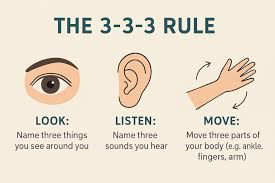
Anxiety and stress reduction
This is an easy method to shift your attention. Begin by naming three objects that you can notice while glancing around. Next, pay attention. Which three noises are you aware of? Next, make three movements with your body: flex and release your shoulder, or move your fingers and toes.
4. Physical activity;
Not an athletic or long- distance runner? its probably not the right time to begin intense training. But keep in mind that exercise in any form is healthy for you and can reduce anxiety symptoms.
Walking, yoga, and tai chi are example of mild activity that releases these feel good chemicals. If you can’t get to those right away, do so me stretches at your desk or go for a quick lunchtime stroll outside.
5. Music;

Anxiety and stress reduction
Soothing music is beneficial for persons with mild or severe anxiety, according to a 2015 study. It has been demonstrated that music lowers blood pressure and heart rate. To simply listen to your favourite songs or even the sounds of nature, keep music on hand.
Summary
Create playlists so you can rapidly experience symptom relief by listening to them. Additionally, research shows that singing lowers anxiety by generating oxytocin and endorphins. It appears that being good is not really necessary. Just sing

Asthma management tips. People with asthma can successfully manage their symptoms, lessen the frequency of exacerbations, and enhance their quality of life by implementing these management techniques into their everyday lives. Long-term asthma control requires constant collaboration with medical professionals and strict adherence to medication regimens.
ASTHMA MANAGEMENT TIPS
A customized, proactive strategy that combines daily controller medication, quick-relief inhalers for flare-ups, and trigger avoidance is necessary for effective asthma management. A doctor’s documented Asthma Action Plan, which outlines when to take medication, when to raise dosages, and when to seek emergency care, is crucial.
1. Medication Adherence:

• Controller Medications: These medications are taken regularly to prevent asthma symptoms and reduce airway inflammation. They include inhaled corticosteroids, long-acting beta-agonists, leukotriene modifiers, and immunomodulators.
• Reliever Medications: Short-acting beta-agonists (SABAs) are used as rescue medication to relieve acute asthma symptoms and provide quick relief during exacerbations.
2. Asthma Action Plan:
• Develop a personalized asthma action plan with your healthcare provider. This plan outlines daily medication use, steps to take when asthma symptoms worsen, and when to seek emergency care.
3. Avoiding Triggers:
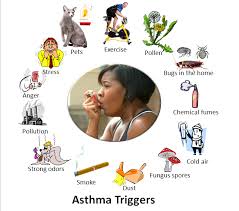
• Identify and minimize exposure to asthma triggers such as allergens (pollen, dust mites, pet dander), air pollution, tobacco smoke, cold air, and respiratory infections.
4. Allergy Management:
• Address underlying allergies that may exacerbate asthma symptoms through allergen avoidance, allergen immunotherapy (allergy shots), or medications such as antihistamines and nasal corticosteroids.
5. Lifestyle Modifications:

Asthma management tips
• Maintain a healthy lifestyle with regular exercise, balanced nutrition, adequate hydration, and sufficient sleep. Exercise-induced asthma can be managed with pre-exercise bronchodilators and warm-up routines.
• Avoid exposure to irritants such as strong odors, chemicals, and air pollutants that can trigger asthma symptoms.
6. Monitoring:
• Use peak flow meters or spirometry to monitor lung function regularly and track asthma symptoms. This helps in early detection of worsening symptoms and adjustment of treatment accordingly.
7. Regular Follow-up:

Asthma management tips
• Schedule regular follow-up appointments with your healthcare provider to assess asthma control, review treatment effectiveness, adjust medication dosages if necessary, and address any concerns or questions.
8. Emergency Preparedness:
• Know how to recognize worsening asthma symptoms and when to seek emergency medical care. Carry a rescue inhaler at all times and ensure family members, caregivers, and school personnel are aware of your asthma action plan.
9. Education and Support:

Asthma management tips
• Educate yourself and your family about asthma management, including medication use, trigger avoidance, and recognizing signs of worsening symptoms. Join support groups or seek counseling to cope with the emotional aspects of living with asthma.
10. Environmental Control:
• Managing asthma requires taking steps to control indoor environmental factors such as dust, mold, and humidity levels. Use mattress and pillow covers, vacuum regularly, and maintain proper ventilation to reduce allergen exposure.
Summary
To control symptoms, avoid exacerbations, and preserve general lung health, asthma management entails a combination of medication, lifestyle modifications, and routine monitoring. The following are some essential asthma control techniques:

Tips for Emotional stress

Anxiety and stress reduction

Face cleansing tips

A Step-by-Step Guide to Deleting Reels on Instagram

A Comprehensive Guide to Setting Up a YouTube Premiere

How to Create a WhatsApp Group
-

 Social media2 weeks ago
How to Delete Reels on IG
-

 Social media2 weeks ago
Tips for likes and Followers on Facebook
-

 Music2 weeks ago
Lyrics for Iké Nilé
-

 Social media2 weeks ago
Guidelines for Creating a Facebook Page
-

 Photography2 weeks ago
Photography and Outdoors
-

 Social media2 weeks ago
Guidelines for Uploading Videos on Youtube