Health
Alcoholic Liver Diseases
ALCOHOLIC LIVER DISEASES
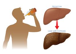
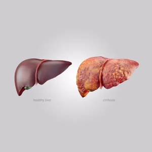
Alcoholic liver diseases. From cirrhosis to fatty liver, alcohol-related liver disease is a collection of liver disorders brought on by excessive alcohol usage. A major worldwide health concern, it contributes to high rates of illness and mortality. Damage to the liver caused by excessive alcohol consumption is known as alcoholic liver disease. It may result in scarring, inflammation, and fat accumulation.
ALCOHOLIC LIVER DISEASES
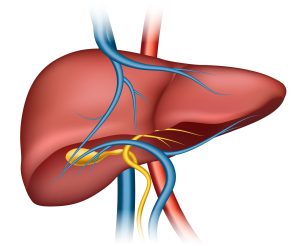
With more than 500 functions, the liver is one of the body’s most intricate organs. These consist of: removing poisons from the blood conserving energy and producing proteins and hormones controlling blood sugar and cholesterol.
Signs
Alcoholic liver disease has hazy early symptoms that impact several bodily systems.
In addition to feeling generally ill, symptoms may include:
pain in the abdomen, nausea, vomiting, and diarrhea
reduced desire for food
Early symptoms can be easily written off as the result of a gastrointestinal ailment or general malaise. However, if these symptoms are not identified and treated, particularly if alcohol consumption persists, liver disease may develop more quickly over time.
Fatty liver disease caused by alcohol
Fatty acid buildup in the liver might result from heavy alcohol consumption. This can occasionally be brought on by substantial drinking over a brief period of time—even less than a week.
If the person stops drinking alcohol after this, alcoholic fatty liver disease is frequently reversible and typically has no symptoms.
Hepatitis caused by alcohol
One severe form of alcoholic liver damage is alcoholic hepatitis. Any cause of liver enlargement and inflammation is referred to as hepatitis.
Continued alcohol consumption will result in persistent inflammation of the liver. After years of heavy drinking, this can happen. Acute episodes of it can also happen during binge drinking episodes.
Fibrosis
A buildup of specific proteins, such as collagen, in the liver is known as fibrosis. It is present in the majority of chronic liver diseases.
Physicians utilize the Metavir grading system, which ranges from A0 to A3, to assess the degree of fibrosis:
A0: no action
A1: minimal activity
A2: moderate level of activity
A3: intense activity
Cirrhosis
Long-term inflammation of the liver causes cirrhosis, which results in scarring and loss of function. This illness has the potential to be fatal. Damage from cirrhosis cannot be reversed, but by continuing to abstain from alcohol, one can stop additional harm. Although liver function can be improved by lifetime abstinence, cirrhosis can cause severe and permanent damage that may require a liver transplant in order to survive.
Treatment
Abstinence
some early stages of liver disease may be reversed as a result. For instance, quitting alcohol after being diagnosed with fatty liver disease may help cure the condition in as little as two to six weeks. A doctor will advise someone with alcoholic liver disease to never drink again once they have been diagnosed at any point. Once drinking resumes, any symptoms that have been reversed will usually return.
Therapy
However, benzodiazepines and cognitive behavioral therapy can help people with alcoholism who are experiencing withdrawal symptoms. For closer supervision, those with severe alcoholism may remain in an inpatient recovery center. After then, continuing treatment could be necessary to avoid relapsing into alcohol consumption. Additionally, medications can stop relapses.
Changes in lifestyle
Also, because smoking and being overweight have both been linked to the deterioration of alcoholic liver disease, doctors may also advise patients to stop smoking and lose weight. Additionally, doctors could advise taking a multivitamin every day.
Summary
The degree of liver damage, the existence of risk factors and comorbidities, and the capacity to fully abstain from alcohol all affect the prognosis for individuals with ALD. Results are often better for people with moderate disease, who have few or no risk factors and problems, and who are abstinent.


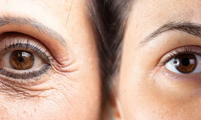
Health benefits and effects of sunlight. Vitamin D production, enhanced mood through serotonin, and balanced sleep cycles are just a few of the vital health advantages of sun exposure. However, excessive UV radiation exposure results in both short-term problems like sunburn and long-term harm including accelerated skin aging (wrinkles, spots), a compromised immune system, and a higher chance of skin cancer.
HEALTH BENEFITS AND EFFECTS OF SUNLIGHT
Sunlight, a celestial orb of energy that has been revered throughout history, is necessary for life to exist on Earth. In addition to providing warmth and illumination, sunlight has several positive effects on human health. In this investigation, we discover the many benefits and profound impacts of embracing the sun’s nourishing rays.
1. Vitamin D Synthesis:

• Effect: Sunlight is a natural source of ultraviolet B (UVB) rays, which trigger the synthesis of vitamin D in the skin.
•Advantages include improved immune system performance, healthier bones, and a lower risk of developing certain diseases when vitamin D levels are adequate.
2. Mood Enhancement:
• Effect: Sunlight exposure stimulates the production of serotonin, a neurotransmitter linked to mood regulation.
• Benefits: Natural light can improve mood, alleviate symptoms of depression, and contribute to an overall sense of well-being.

3. Regulated Circadian Rhythms:
• Effect: Exposure to natural light helps regulate the body’s internal clock, known as the circadian rhythm.
• Benefits: Maintaining a healthy circadian rhythm enhances sleep quality, mood stability, and overall physiological function.
4. Improved Sleep Quality:

• Effect: Exposure to sunlight, particularly in the morning, helps signal to the body that it is daytime, promoting wakefulness.
• Benefits: Regular exposure to natural light contributes to a more robust sleep-wake cycle, leading to improved sleep quality.
5. Enhanced Skin Health:
• Effect: Sunlight exposure has positive effects on certain skin conditions, such as psoriasis, acne, and eczema.
• Benefits: Moderate sun exposure can promote skin healing and alleviate symptoms of various skin conditions.
6. Boosted Immune Function:

• Effect: Vitamin D, produced through sunlight exposure, plays a crucial role in supporting a robust immune system.
Benefits: Having enough vitamin D has been related to a lower risk of infections and autoimmune diseases.
7. Cardiovascular Health:
• Effect: Sunlight exposure is linked to the production of nitric oxide, which can help regulate blood pressure.
• Benefits: Improved cardiovascular function and reduced risk of heart-related issues.
8. Cancer Prevention:

HEALTH BENEFITS AND EFFECTS OF SUNLIGHT
• Effect: Adequate vitamin D levels, synthesized through sunlight exposure, have been associated with a reduced risk of certain cancers.
• Benefits: Sunlight may contribute to cancer prevention, particularly for cancers of the colon, breast, and prostate.
9. Bone Health:
• Effect: Vitamin D, facilitated by sunlight, is essential for calcium absorption and bone health.
Adequate vitamin D levels contribute to strong and healthy bones, reducing the risk of osteoporosis.
10. Positive Impact on Mental Health:

HEALTH BENEFITS AND EFFECTS OF SUNLIGHT
Sunlight exposure has been linked to reduced symptoms of Seasonal Affective Disorder (SAD) and other mental health conditions.
– Benefits: Natural light can uplift mood, reduce stress, and contribute to a positive mental state.

HEALTH BENEFITS AND EFFECTS OF SUNLIGHT
The sun provides numerous health benefits beyond just warmth and light; it is frequently lauded as a symbol of vitality and life. Embracing the sun responsibly—mindful of potential risks such as sunburn and skin cancer—can be a powerful and natural way to enhance physical and mental well-being. As we bask in the rays, let’s appreciate the intricate dance between sunlight and our bodies, a harmonious symphony that orchestrates health and vitality. Read more articles here.
Summary
The body can synthesize vitamin D and other non-vitamin D routes, like the synthesis of serotonin and nitric oxide, when exposed to moderate amounts of sunlight. However, there are serious hazards associated with excessive exposure, such as early skin aging and skin cancer.

Optimal health and water consumption. Daily hydration is necessary for optimal health in order to support vital processes like waste elimination, joint cushioning, and temperature regulation. Although the “8×8 rule” (eight 8-ounce glasses) is a well-liked, simple-to-remember objective, typical guidelines are about 11.5 cups (2.7 liters) per day for women and 15.5 cups (3.7 liters) per day for men, including water from all foods and drinks.
OPTIMAL HEALTH AND WATER CONSUMPTION
The recommended daily intake of water from all foods and beverages is about 2.7 liters (11.5 cups) for women and 3.7 liters (15.5 cups) for men. Sufficient consumption is essential for physiological processes such as controlling body temperature, eliminating waste, and staying hydrated. Activity, heat, and medical problems all raise needs.
1. Fundamental Hydration:

It serves as the foundation for staying hydrated, a fundamental aspect of maintaining bodily functions. Adequate hydration is vital for supporting cellular processes, regulating body temperature, and ensuring the optimal function of organs and systems.
2. Nutrient Transportation:
Water acts as a universal solvent, facilitating the transportation of essential nutrients throughout the body. It aids in the digestion, absorption, and distribution of nutrients from the food we consume, ensuring that our cells receive the necessary fuel for energy production and overall vitality.
3. Detoxification and Waste Removal:

The human body constantly produces waste products as a result of metabolic processes. Water plays a pivotal role in flushing out these toxins through urine, sweat, and other bodily fluids. Proper hydration supports the efficient removal of waste, promoting a healthy and functioning internal environment.
4. Joint Lubrication:
Water is a key component of synovial fluid, which lubricates joints and provides cushioning for bones. Maintaining adequate hydration is essential for joint health, as it helps reduce friction between bones and supports smooth, pain-free movement.
5. Temperature Regulation:

As the body heats up during physical activity or in warm environments, water plays a crucial role in cooling through processes like sweating and evaporation. Proper hydration is essential for effective temperature regulation, preventing overheating and maintaining optimal physiological balance.
6. Cognitive Function:
Dehydration can negatively impact cognitive function, leading to decreased alertness, concentration, and short-term memory. Ensuring sufficient water intake is vital for maintaining optimal brain function and supporting mental clarity
7. Skin Health:

OPTIMAL HEALTH AND WATER CONSUMPTION
Water is vital for skin hydration and elasticity. Proper hydration contributes to a healthy complexion, reducing the likelihood of dryness, flakiness, and premature aging. Drinking enough water supports the body’s natural ability to maintain skin health frm the inside out.
8. Cardiovascular Support:
Adequate hydration is crucial for maintaining blood volume and supporting cardiovascular health. Proper blood circulation ensures that oxygen and nutrients are efficiently transported to cells, while waste products are removed, contributing to overall heart health.
9. Muscle Function:

OPTIMAL HEALTH AND WATER CONSUMPTION
Staying hydrated is essential for muscle contraction and energy production during physical activity. Dehydration can lead to muscle cramps, fatigue, and decreased exercise performance. Optimal hydration supports muscle function, endurance, and recovery.
10. Weight Management Aid:

OPTIMAL HEALTH AND WATER CONSUMPTION
Drinking water before meals can contribute to a feeling of fullness, potentially reducing overall calorie intake and supporting weight management efforts. Staying hydrated can also prevent confusion between thirst and hunger cues. .Stay tuned for more updates.
Summary
An essential part of our existence is water, the elixir of life. Beyond only keeping us hydrated, water is essential for many other aspects of our general health and wellbeing. In this investigation, we examine the vitality of water and its significant influence on how our bodies operate.

Analysing nostalgia. A nostalgic yearning for the past is the hallmark of nostalgia, a complicated, bittersweet emotion that is frequently brought on by sensory cues like music, fragrances, or sentimental mementos. Although it was long thought of as a sickness, contemporary research shows that it is a healthy, social feeling that improves mood, self-esteem, and fortifies social bonds during periods of transition or loneliness.
ANALYSING NOSTALGIA
As a psychological resource, nostalgia improves mood, fortifies social ties, raises self-esteem, and builds resilience against loneliness or life changes. Although it typically encourages optimism and a sense of purpose in life, it can sometimes cause melancholy or impede emotional healing if it results in excessive reflection on an idealized past.
What is Nostalgia?

Nostalgia is the happy feeling you get when you remember fun and exciting things from a long time ago. It’s like having a treasure chest full of special memories, and each memory is like a little treasure that makes your heart feel good.
Imagining a Time Machine:
Think of nostalgia as a magical time machine. When you close your eyes and think about your favorite moments, like playing with friends, celebrating birthdays, or cuddling with your favorite stuffed animal, it’s like taking a ride in this special time machineExploring Memories:
Nostalgia is like opening a storybook filled with pictures of the past. Each page holds a different adventure, like the time you learned to ride a bike or the day you met your best friend. These memories are like colorful puzzle pieces that make up the picture of your life.
Why is Nostalgia Important?

ANALYSING NOSTALGIA
Nostalgia is important because it helps us remember the good things in life. When we feel a little sad or miss someone, thinking about happy memories can make us feel better. It’s like having a superpower that turns frowns into smiles.
Sharing Nostalgia:

ANALYSING NOSTALGIA
You can share the memories with others by talking about your favorite memories. When you share, it’s like giving a piece of your happy feelings to someone else. You can say, “Remember that time we built a sandcastle at the beach?” and watch as everyone smiles together.
Nostalgia is a special friend that brings joy to our hearts. It’s not about living in the past but cherishing the wonderful moments that shape who we are. So, next time you feel that warm and fuzzy feeling, know that you’re experiencing the magic – your very own time machine of happiness. Stay tuned for more updates.
Summary
A wonderful emotion that transports us back in time is nostalgia. We smile as it envelops us in a warm, fuzzy blanket of memories. It may sound like a huge term, but let’s take a closer look at what makes it so unique.

Airline Travellers and Baggage Allowances

First Time Travellers and Airline Policies

Plane Tickets Relevance

A Step-by-Step Guide to Deleting Reels on Instagram

How to Create a WhatsApp Group