
Health
Treatments for Ear Infections
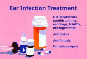
TREATMENTS FOR EAR INFECTIONS
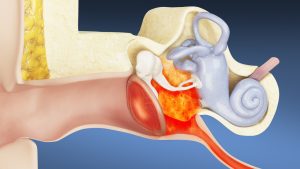
Treatments for ear infections. Both bacteria and viruses can cause ear infections. They can happen in the outer and inner ear, as well as the middle ear, which is the area of the ear directly behind the eardrum. Although they frequently go away on their own, swelling or fluid accumulation can make them painful. Both acute and chronic ear infections are possible. Although they hurt, acute ear infections don’t last long. Ear infections that are chronic either don’t go away or keep coming back. They may result in middle and inner ear damage, which is rarely irreversible.
TREATMENTS FOR EAR INFECTIONS
To diagnose an ear infection, a doctor frequently only needs a device known as a pneumatic otoscope. By using this tool, the physician can examine the ear and determine whether fluid is present behind the eardrum. The doctor gently presses air against the eardrum using the pneumatic otoscope. Normally, the eardrum would move in response to this puff of air. Your doctor will notice little to no movement of the eardrum if the middle ear is filled with fluid.
Treatment at home

The following techniques work well for reducing the signs of a minor ear infection:
• Place a warm cloth over the ear that is afflicted.
• Use over-the-counter pain relievers like Tylenol’s acetaminophen or ibuprofen.
• To relieve pain, apply over-the-counter or prescription ear drops. Decongestants such as pseudoephedrine are available over-the-counter.
• Do not sleep on the ear that is afflicted.
Medical care
Also, consult a physician if your symptoms worsen or remain the same. If your ear infection is bacterial, persistent, or doesn’t seem to be getting better, they might recommend antibiotics. Viral infections cannot be treated with antibiotics.
Children’s medical care

Meanwhile, in order to prevent overprescribing antibiotics, which can result in antibiotic resistance, doctors frequently treat pediatric ear infections by waiting and seeing. If your symptoms are severe or don’t go away in two to three days, your doctor might occasionally prescribe antibiotics. As an alternative, they might write you a prescription but advise you to wait two to three days to see if your child’s symptoms improve. Children should never take aspirin without a doctor’s prescription. One avoidable risk factor for Reyes’ syndrome, a rare condition that damages the liver and brain, is taking aspirin.
Surgery

Although, If the standard medical treatments don’t clear up your ear infection or if you get ear infections frequently in a short period of time, surgery might be your best bet. Ear tubes are typically inserted into your ears to let fluid escape. Your eardrums are surgically opened to accommodate these tubes. The holes eventually heal after they fall out. Occasionally, surgery is required to close these holes.
Prevention
The following behaviors may lower your risk of developing an ear infection:
1. frequent hand washing
2. avoiding crowded places
3. not using pacifiers around babies and young children;
4. breastfeeding babies; avoiding secondhand smoke
5. maintaining current vaccinations
The bacteria or viruses that cause ear infections are found in the middle ear, which is the area of the ear behind the eardrum. The majority of ear infections go away in three days or less, but more serious infections might require antibiotic treatment. Children are most likely to get ear infections. If you or your child experience severe pain, a fever exceeding 102.2°F, ear drainage, or other worrisome symptoms, it’s imperative that you consult a physician.
Summary
A virus or bacteria that infects the area behind your child’s eardrum can cause ear infections, also known as acute otitis media. One of the symptoms is ear pain, which can make your baby or toddler particularly fussy or agitated. Ear infections frequently go away on their own. Children occasionally require ear tubes, antibiotics, or painkillers.


Cardiac arrest prevention tips. A abrupt loss of heart function, or cardiac arrest, occurs when the heart ceases to beat efficiently. A heart attack, an irregular heartbeat, or other underlying medical issues are some of the possible causes. Nonetheless, there are steps you may take to avoid cardiac arrest. Getting medical help right once is essential to restoring the heart’s regular rhythm and function. It’s a major medical emergency, and survival rates can be significantly increased with early intervention, such as CPR and the use of a defibrillator.
CARDIAC ARREST PREVENTION TIPS
A abrupt, unanticipated loss of heart function brought on by an electrical fault in the heart is known as cardiac arrest. It causes the person to collapse, lose consciousness, and cease breathing regularly because it prevents blood from reaching the brain and other organs. It is a medical emergency that, if left untreated, can be lethal in a matter of minutes.
Building the foundation;

Cardiac arrest prevention tips
Establishing heart-healthy habits early is paramount. Emphasizing a diet rich in fiber, fruits, vegetables, and lean proteins, coupled with regular physical activity, sets the foundation for a healthy heart. Stress-free environments and good sleep hygiene in childhood contribute to reducing heart disease risks later in life.
Healthy diet guidelines for young adults;

Maintaining a balanced diet in your 20s and 30s involves monitoring saturated and trans fats, choosing healthier fat sources, and being cautious about added sugars. Reading food labels, opting for whole, unprocessed foods, and customizing dietary choices contribute significantly to heart health. Quitting smoking and vaping is a transformative step for heart health. It reduces the risk of heart attacks, normalizes blood pressure and heart rate, improves blood flow, decreases inflammation, and enhances overall cardiovascular function. The benefits extend beyond the individual, protecting loved ones from secondhand smoke.
Maintaining vigilance;

In your 20s and 30s, when distractions abound, sustaining healthy habits becomes challenging. Reducing screen time, including physical activity, and adopting a heart-friendly diet are crucial. Avoiding smoking and vaping, managing stress, and customizing an exercise plan based on individual preferences and health conditions are vital steps.
Routine check-ups in your 40s and 50s;

As your age adds, routine check-ups become imperative. Tests like blood pressure screening, cholesterol profile, blood sugar tests, and EKGs help identify potential risks. Embracing a healthy routine, staying vigilant for symptoms, and seeking prompt medical attention are essential steps in maintaining heart health
Summary
Managing underlying cardiac problems and implementing heart-healthy lifestyle practices are necessary to prevent cardiac arrest. Keeping your heart and blood arteries in top condition is the key to reducing your risk because coronary artery disease is a major contributing factor.

Sleep and long term health. One of the most important pillars of physical wellness is getting enough good sleep. It enables self-healing and self-regulation of your immune, endocrine, and cardiovascular systems. Chronic sleep deprivation raises the long-term risk of serious illnesses like obesity, diabetes, and heart disease.
SLEEP AND LONG TERM HEALTH
Just as important as nutrition and exercise is getting enough sleep. Your long-term risk of serious medical illnesses, such as type 2 diabetes, cardiovascular diseases, and neurodegenerative disorders, is greatly increased by chronic sleep deprivation (sleeping fewer than 7 to 9 hours every night). Sleeping enough at night shields you against a host of health issues. Here are a few of the most significant ones.
Heart disease

A healthy sleep pattern lowers the risk of heart disease. In the United States, it is the main cause of death for both men and women. Your blood pressure and heart rate decrease as you sleep. Your heart will have to exert less effort as a result.3. Sleeping well also contributes to a healthy cortisol balance. If this stress hormone remains too high for an extended length of time, it might be harmful to the body.
Weight gain
Increasing your sleep will assist you in managing your weight. Your body produces an excess of a chemical that causes hunger when you’re sleep deprived. What was the outcome? You consume more food than you usually do. And you have a craving for foods heavy in sugar and fat. Maintaining a healthy weight reduces your chances of high blood pressure, high cholesterol, and other conditions.
Best advice for a great night’s sleep;

You can now see why getting more sleep is necessary. These hints will improve your quality of sleep at night.
Stick to a schedule
Every day, get up and go to bed at the same hour. On the weekends, it could be tempting to stay up late and sleep in. However, that simply makes Monday morning more difficult for you to wake up.
Don’t eat too late

Sleep and long term health
Heartburn can occur when a full meal is consumed close to bedtime. Chest discomfort that burns is one of the signs of heartburn. This may prevent you from going to sleep and remaining asleep. Before going to bed, eat something small and light, like a banana, if you’re hungry. Alternately, have a low-sugar yogurt cup or some almonds.
Nap wisely
If you must nap throughout the day, try to limit the duration to no more than 20 minutes and take it before 3 p.m. Sleeping at night becomes more difficult after extended, late naps.
Cut down on screen time
Melatonin is disrupted by the blue light emitted by your computer, tablet, and phone screens. This is a hormone that makes you feel drowsy by rising naturally around bedtime. When the light is too strong, your body suppresses melatonin because it believes it is still daytime. At least half an hour before going to bed, turn off all gadgets.
Give your bedroom a makeover

Sleep and long term health
Make a relaxing, peaceful sleeping area. Turn down the lights before going to bed, and avoid sleeping with the TV on. To make your room as dark as possible, hang curtains or room-darkening shades on your windows. To reduce noise, use a free white noise app on your phone. Moreover, maintain a temperature of between 60° and 67°F.
Limit fluids
If you have too much water right before bed, you’ll wake up in the middle of the night to go to the bathroom.
Summary
Consistent lifestyle choices that avoid chronic illness and preserve cognitive function are essential for long-term health. A healthy, plant-based diet, 150 minutes of aerobic exercise plus strength training, getting enough sleep, and abstaining from smoking are all important practices.

Nivea body wash for men

Business maintenance

Athlete conviction of murder

A Step-by-Step Guide to Deleting Reels on Instagram

A Comprehensive Guide to Setting Up a YouTube Premiere

How to Create a WhatsApp Group
-

 Health4 weeks ago
Carbonated drinks side effects
-

 Business4 weeks ago
Smart ideas for business startups
-

 Deodorants3 weeks ago
Reviewing tom ford oud
-

 Deodorants4 weeks ago
Reasons for wearing deodorants
-

 Deodorants4 weeks ago
Review for saint laurent black
-

 Deodorants3 weeks ago
Varieties of perfumes for men